


Resolving Power (Part 1)
How technology unlocks the past
Risk & Progress| A hub for essays that explore risk, human progress, and your potential. My mission is to educate, inspire, and invest in concepts that promote a better future. Subscriptions are free, paid subscribers gain access to the full archive, including the Pathways of Progress and Realize essay series.
We are but brains on sticks. Our bodies are extensions of the brain, gathering sustenance and providing sensory input so “we” can interact with the physical world. But our ability to sense and understand the universe is inherently restricted by our physical limitations. Our eyes can only perceive a narrow band of wavelengths in the electromagnetic spectrum, and our resolving power is just as limited. Technological advancements, the fruits of human progress, have made it possible to extend our senses, to study and observe a hidden world, from the smallest particles to the most distant objects.
A Hidden World
The naked human eye can resolve objects about as small as 100um or the width of a human hair. For most of our history, this was the limit. There is evidence, however, that some ancient societies may have used polished transparent rocks as crude ‘lenses’ thousands of years ago. One of the earliest examples is the “Nimrud lens” dating to around 750 BC, though there is some debate as to whether or not it was a decorative piece or used for practical purposes. These early lenses’ primary use may have been to focus sunlight to create fire, though conceivably, they could have also been used to magnify small objects.
The earliest glass lenses explicitly designed for magnification emerged in Italy in the late 1200s or early 1300s, produced by spectacle craftsmen. These early lenses had a resolving power of 6-10x at most. The first attempt at a “compound microscope,” which utilized multiple lenses inside a tube to enhance magnification, is often attributed to Dutch craftsman Zacharias Janssen and his father Hans around 1590. These early microscopes were limited by the quality of optics available at the time, reaching a maximum magnification power of 30-40x in the following decades.
In 1675, the “father of microscopy,” Anton van Leeuwenhoek, found that he could increase the magnification power of lenses by grinding and polishing them with greater precision. His simple, single-lens ‘microscope’ could magnify images up to 270x. With it, Leeuwenhoek was the first human to see bacteria, red blood cells, muscle tissue, and the microscopic world beneath and around us. He also witnessed life teeming inside what otherwise appeared to be clear water droplets. His study of this microscopic world laid the groundwork for the germ theory of disease that would be confirmed two centuries later and save millions of lives.
Leeuwenhoek’s microscope was better described as a high-powered magnifying glass. Compound microscopes had a lot more potential but were still limited by issues with light diffraction (chromatic aberration) and image blurring (spherical aberration). In 1729, Chester Moore Hall presented the first achromatic lens that greatly reduced chromatic aberration. Over the next century, improved, high-purity glass and better manufacturing techniques led to still better image quality. Then, in 1830, Joseph Jackson Lister was the first to describe a chromatically and spherically corrected compound microscope. Around this time, mirrors were added to microscopes to gather more light, consequently, their resolving power greatly increased.
The best light microscopes, however, were now limited by the wavelength size of visible light. They could magnify up to 1000x, resolving objects about as small as 200nm or the size of some organelles that comprise individual cells. To see objects any smaller, we had to ditch visible light itself. It didn’t take long to figure out how. The electron microscope was born just a century after “perfecting” the visible light microscope. The electron microscope uses a beam of electrons instead of visible light to interact with the objects being studied, focused by electromagnetic lenses instead of glass.
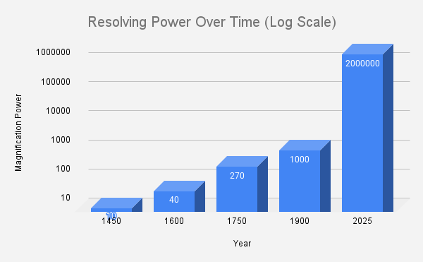
Because electrons are particles and much smaller than light waves, we could dramatically increase the resolving power of the human eye, initially magnifying objects up to 200,000x and, as they improved, 2,000,000x. This enabled the study of molecules and even to “see” individual atoms as small as 1nm across. For the first time, we could see and study DNA, the blueprints of life, we could understand small cellular structures, detect and study viruses that kill, and examine the fine details of materials to improve our materials science.
Just as the electron microscope was adopted, however, physicists began to understand that a “subatomic” world existed at scales smaller than the atom. Studying this world required another medium shift, another technological breakthrough, and it was just around the corner: particle accelerators. By accelerating subatomic particles like protons or electrons at high speed, controlled and guided by electromagnetic fields. Particle accelerators aided in the study and discovery of subatomic particles, the forces of nature, and have helped us understand the universe at its most fundamental level.
Peering into the Past
Our ability to resolve and study ever smaller objects has metaphorically enabled us to peer back in time. Our eyes can only see the natural world as it is, but light microscopes reveal the world of cells and bacteria, helping unlock the mysteries of biological evolution. Electron microscopes went further back, unveiling the atomic world and helping shed light on chemical evolution. Particle accelerators went further still, aiding in understanding the fundamental laws of physics that were born in the first moments of the universe, taking us back 14 billion years.
You also may like….

